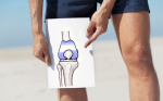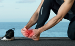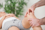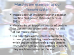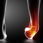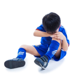
Knee Pain
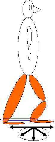
Have you considered the control over the knee occurring from ball & socket joints? Traditionally, anatomical constructs of reasoning suggest that the lateral & medial stability of the knee is precariously maintained by collateral ligaments. However, what controls the tibia and femur? Since, the hip and talo-navicular joints are ball & socket joints they serve the function of providing ROM. The motor control conundrum that such large ROM presents is it's balance with stability not only in the hip and talonavicular joint but also those regions abilities to contribute to the protection of the ligaments of the knee.

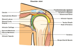
link to explanation in Shoulder section
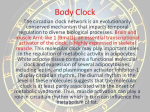
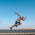
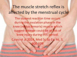
Motor learning theory devised by Bernstein suggests that the body will use the momentum of the limbs to optimize the degrees of freedom in the system. Similar to a mass-spring analogy where the perturbations of the mass will be dependent on the damping characteristics of the spring, the brain will introduce muscle tone to dampen the angular velocity and hence acceleration of the system. Therefore, instead of the muscles of the leg lifting the limb, the antagonistic muscles are decelerating the limb towards the end of trajectory. Moreover, by using eccentric (muscle lengthening and contracting) muscle contractions the system becomes efficient through these decelerating movements through enhanced visco-elastic rebound as well as the conservation of momentum.
see Motor Learning section for further explanations
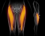
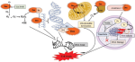
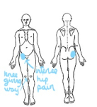
As such, the muscles around the hip and talo-navicular joints perform the function of multidimensional stability. Clearly, the femur affects knee positioning and the position of the ilium/hip will affect timing of the muscles around the hip-thigh. Any anterior ilial rotation generally makes it difficult for the gluteus maximus to fire before or simultaneously with the hamstring muscles. Tightness of the adductor muscle can cause an 'inflare' of the ilium, potentially placing adverse tension on the ischiococcygeal and sacrotuberal ligaments. Moreover, this adverse tension of the adductors could affect the nutrition to the knee through the phylogenetic link of these muscles with the medial collateral ligament and hence medial meniscus. Any adverse function of the iliopsoas can affect femoral blood flow which is likely to affect slow twitch, stabilising, endurance muscles more than fast twitch, glycolitic, ballistic muscles. Hence, as the duration of activity increases, movement stability may break down, leading to 'poor form' and potential injury.
The subjective examination should include aspects of the stage, stability, irritability and severity of the disorder.

Hence the physical examination should include analysis of
- gait
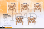
- pelvc symmetry
- lumbo-pelvic dynamics
- femoral pulse
- inferior lateral movements of the T/S and lateral diaphragm control over Psoas Major function

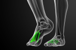
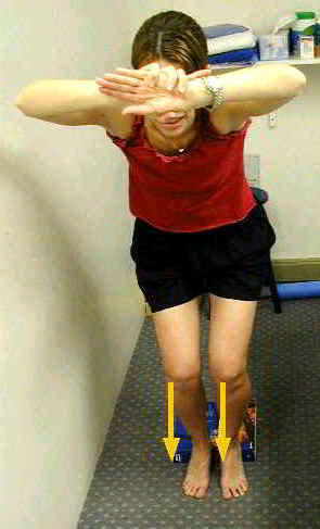
- foot dynamics (esp. pronation supination)
go to foot - orthotics section for further details
- muscle timing and duration of contribution between Vastus Medialis and Vastus Lateralis, Gluteus Maximus and Hamstrings, Gluteus Medius and Adductor synergy, deep hip rotator endurance and strength.
- Relationship between the deep and superficial abdominal muscles and their affect on pelvic symmetry and lumbopelvic rhythm.
- ROM's of the hip, knee, foot and L/S
- Muscle energy techniques
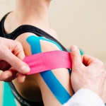
- Taping of the patella
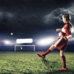
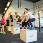
- fit for skiing?
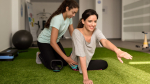
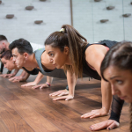
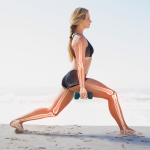
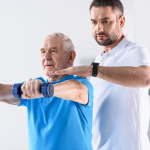
Abdominal and Gluteal Strengthening

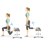
Left Quadriceps Strengthening

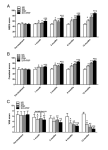
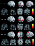
additionally, EMG can be used in combination with transverse abdominis, gluteus medius, medial calf muscles and VMO
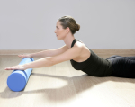
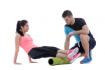
Iliotibial Band lengthening and combined gluteal/quads/abdominal strengthening

Treatment should include techniques and exercises which ameliorate the problems identified during the examination. These may include muscle energy techniques (MET), soft tissue massage (STM), dry needling (intramuscular stimulation {IMS}), EMG biofeedback, taping, joint mobilisations, and motor learning principles.
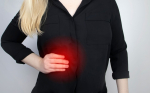
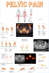
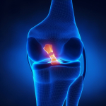
Patella Tracking Dysfunction
Similarly, you may have to consider the influence of the pelvis in patella tracking dysfunction. If the pelvis has an adductor longus with high tone then this could rotate the hemi-pelvis inwards on the stance leg. Subsequently tension is generated in the lateral rotators of the hip (eg obturator internis and externis) which could irritate the obturator nerve creating further spasms in the adductor longus. This in turn may generate shearing across the pubic symphysis resulting in pain in the groin. Additionally, the rotation &/or inflare of the hemipelvis may result in anterior ilial rotation (counter-nutation) creating an irritated rectus femoris and ITB/Tfl. Furthermore, this counternutation may place excessive strain through the biceps femoris resulting in sacrotuberal and sacroiliac ligament irritation.

Taping or other aids can also be used around the hip/buttocks to prevent femur internal rotation thereby reducing the relative lateral alignment of the patella w.r.t the femur.
In conclusion, the examination and treatment of the knee should include assessment of all the structures which can affect knee function. Prior to the physical examination, a thorough subjective examination should be conduct to enable the client and the therapist to engage in the clinical reasoning process. Interested readers should look at the Instructional Design section of this website.

Last update : 3 September 2012
 What We do
 What We Treat