Clinical Physiotherapeutic Clinical Reasoning in Assessing Structural Instability
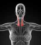
Upper cervical instability although rare, can be catastrophic and shouldn't be missed by a physiotherapist. On the few occassions where I have come across Upper Cervical Spine instability, there was severe restriction of movement due to protective muscle spasms. The nature of these type of muscle spasms is quite different to that of functional instability or painful dysfunction. During manual therapy, generally speaking muscle spasms reduce gradually over a 15 -30 second period during joint mobilisations of a structurally intact cervical spine. However, where the integrity of bones or ligamentous support is compromised, the nature of muscle spasms can be extremely severe masking the nature of the instabilty. In other cases, where partial catastrophic failure and neural destruction has already occured the symptoms are more obvious. Again where intermittent paralysis and/or reflexogenic inhibition results voluntary restriction of ADL , positive Instability Testing usually confirms diagnosis.
The 'classic' case is from football (soccer) history. One of Bert Trautmann's greatest matches was the legendary 1956 FA Cup Final between Manchester City and Birmingham City at Wembley Stadium. In the 75th minute Man City led 3:1 and Trautmann, diving at an incoming ball, was knocked out in a collision with a Birmingham attacker when he was hit in the neck. For the remaining 15 minutes he defended his net, because at the time there were no substitutions possible. Manchester City held on for the victory, and the hero of the final was Bert Trautmann, due to his spectacular saves in the last minutes of the match. Three days later, an x-ray revealed he had a broken vertebra in his neck.
In several cases which I have come across, diagnosis was not immediate due to the intermittent nature of neural compromise and/or due to the severity of movement restriction from muscle spasms.
In one case, a truck driver had his several tonne load fall on him. He drove his truck to the hospital where he was given a mild painkiller and told to take it easy. He drove from Port Augusta back to Lithgow (approx 2000km) and then took 6 weeks off work feeling pretty miserable. Fortunately, a friend visited him who happened to be a doctor, who drove him to the local hospital where he insisted on correct imaging which resulted in an air-ambulance evacuation and emergency surgery (1998).
In another case, a man was walking with his wife one evening when he stepped onto a covered man-whole which gave way and he fell 3m into a shaft approx 1.5m wide. As he landed on the bottom of the shaft, his knees smashed against the wall on one side and his back hit the opposite side. After 1 year of 'severe neck stiffness' a specialist finally confirmed the diagnosis and immediate surgery was performed (1988).
Final case was a bus driver whose bus fell off a road during a landslide. He saved several children and ran some 15km back to a farm to get help. When the local emergency services were slow in coming, he ran back to the bus, where his wife was looking after the surviving children. At the end of the day, the police finally arrived to find the carnage. Eventually, everyone was evacuated to a local hospital and later some were flown out to larger hospitals. When the adrenalin finally wore off, he started to feel pretty bad. When he stated this to the medical officers, they took some X-rays and made him a cup of tea and stated he would be O.K. He then rang an Intensivist medical friend of his, who drove 180km to pick him up and drove the equivalent back again, where his hospital detected 5 vertebral fractures, including a dens fracture (1992).
Certain people may be predisposed to upper cervical spine instability. These people include those suffering from soft tissue hypermobility, joint hypermobility syndrome (JHS), connective tissue disorder (CTD), Marfans Syndrome, Ehler Danlos syndrome (EDS). Dural laxity, vascular irregularities and ligamentous laxity with or without Arnold Chiari Malformations may be accompanied by symptoms of intracranial hypotension, POTS (postural orthostatic tachycardia syndrome), dysautonomia, suboccipital "Coat Hanger" headaches (Martin & Neilson 2014 Headaches, September, 1403-1411). These manifestations need to be borne in mind as not all upper cervical spine instabilities are the result of trauma.
Other examples were people who presented to the practice post-whiplash injury, another who had a manipulation of the upper cervical spine and 2 who had systemic inflammation (Rheumatoid Arthritis, Psoaratic Arthritis) and 1 with a chronic upper respiratory tract infection.
Frequently, physiotherapists rely on several tests to examine the integrity of the dens, transverse and alar ligaments. These tests include
- Sharp Purser (transverse ligament)
- Modified Sharp Purser (transverse ligament)
- Anterior Shear Test (transverse ligament)
- Lateral stability of the A-A joint (0/C1/2) (alar ligament)
- Cervical Distraction with/without flexion (tectorial membrane), with/without rotation, or lateral flexion (alar ligaments)
Functional X-rays include
- through-the-mouth in lateral flexion
- lateral in flexion and extension
These are only useful if the muscle spasm allows sufficient movement to demonstrate excessive gapping b/n the dens and C1 (transverse ligament), or abutting of the dens against C1 in lateral flexion (alar ligaments). Additionally, lack of lordosis may indicate severe muscle spasms.
Remember, the usual MRI of the C/S are taken from C3 - C7, so if you suspect Upper C/S instability then a special request may need to be made.
More importantly, the subjective examination gives decisive information (yellow and red flags) which may make your clinical reasoning skills hone in on Upper Cervical Spine instability. These may include
- mechanism of injury - whiplash, other unguarded impacts, and severe loading can all be yellow flags
- altered sensation and movement of the tongue eg frequent biting of the tongue with potentially little pain
- ataxia
- tinnitus
- diplopia
- dizziness
- dysarthria
- dysphagia
- facial pain/pareasthesia
- feeling of the head 'falling off' when going over a bump in a car
- needing to hold head when getting up out of bed
- neck pain
- headache
- hypoaesthesia of both hands and/or feet
- limb muscle wasting and weakness
- nausea or vomitting
- occipital pain/paraesthesia
- altered sphincter control
- paraesthesia of lips
- retro-occular pain
- spasticity (cord like symptoms)
Physical findings may include positive stability testing, hyper-reflexia in the limbs, babinski, clonus, weakness, loss of balance, loss of active movement due to severe muscle spasms, excessive movement and 'drop attacks' or other unusual neurological symptoms.
People susceptible to a hightened incidence of Upper Cervical Spine instability include those who have
- Sero-negative (don't forget the dandruff) or sero-positive spondylo-arthropathies
- Downs syndrome
- Klippel-Feil syndrome
- Recurrent pharyngeal infections
- Recurrent tonsillitis
- Septic arthritis
- Lupus Erythematosus
- Whiplash or other cervical traumas
This list is by no means exhaustive and it is possible to have some of these symptoms and a totally normal cervical spine. However, listen to the client and take the totality of the examination findings to guide in diagnostic decision making. Importantly, listening may save a persons life from a catastrophic spinal cord injury.
Upper Cervical Spine instability considerations
Aspinall, W., (1990). Clinical Testing for the Craniovertebral Hypermobility Syndrome. JOSPT 12: 2, 47-53.
In occipitalization of the atlas, movement between the occiput and the atlas is abolished. On maximum flexion of the atlas on the axis there is maximal stress on the occipitoodontoid ligaments. Any attempts at lateral flexion or rotation will exert abnormal stress on the occipitoodontoid ligaments with overstretching. This may lead to hypermobility of the atlas on axis with the possibility of atlantoaxial subluxation, which may be found later in life with this type of anomaly or hypomobility of the atlantooccipital joint.
Frequent occurrence of congenital fusion of c2-c3 has been noted in patients with atlantooccipital joint dysfunction. Congenital anomalies of the odontoid or lax transverse ligaments as in Down's Syndrome.
Three types of occipital condyle fractures
type I impacted; ipsilateral alar ligament may be functionally inadequate, producing hypermobility. Stability is maintained by the intact tectorial membrane and the contralateral alar ligament.
type II # into the base of the skull; exists occipital condyle and enters the foramen magnum. Stability is maintained by the intact alar ligaments and the tectorial membrane.
type III avulsion of the occipital condyle by the alar ligament. The contralateral alar ligament and tectorial membrane are loaded, creating a potentially unstable injury. These fractures may be suspected post-trauma if symptoms persist and the patients were initially unconscious on admission, or in those with cranial nerve damage.
Non-union fractures of the odontoid even if initially undisplaced, frequently become displaced.
In atlanto-axial dislocation the atlas slides anteriorly . In patients with pharyngitis and rheumatoid arthritis, the inflammatory process produces pathological laxity of the transverse ligament. The lymphatic drainage of this joint is, primarily, into the retropharyngeal glands which also drain the nasopharynx and ultimately, into the deep cervical glands. Disruption of this lymphatic drainage may have an effect on the laxity of the ligaments.
Rotational fixation of the atlantoaxial joints results in persistent asymmetry of the odontoid in relation to the articular masses of the atlas. This may result in persistent strain on the ligaments and tectorial membrane causing hypermobility. Usual aetiological factors are associated with trauma, but the onset may be spontaneous or associated with upper respiratory tract infection.
the anteroposterior diameter of the ring of the atlas is approx. 3cm, the cord and the odontoid each occupy 1cm, with the remaining 1cm being a potential space.
central cord injury may be found showing greater involvement of the upper as compared to the lower extremities. Motor deficit is more profound than sensory and frequent bladder and bowel dysfunction are often found.
anterior spinal cord injury may develop, with complete paralysis with hypesthesia and hypalgesia to the level of injury. Posterior column functions are relatively spared and the more peripheral column functions such as pain, temperature, and perception are compromised to a lesser degree. Brown-Sequard syndrome may be produced due to hemisection of the spinal cord from a rotational strain.
cord signs -delayed myelopathy ranging from paraparesis to Brown-Sequard syndrome.
- dysaesthesia in the hands, with clumsiness and weakness of hand movements, spastic weakness of the lower limbs, with slight general wasting, and hyperreflexia.
-ankle clonus is sometimes present and the plantar reflexes are likely to be extensor.
-patient will often report difficulty in walking, and sphincter control may be affected.
patients with occipitoatlantial fusion may present with a low hairline and short neck, similar to Klippel-Feil syndrome.
marked inability to push the chin up or press it down against resistance should raise the suspicion of craniovertebral hypermobility.
the alar ligaments could be irreversibly stretched after trauma as they consist of inelastic collagen fibres, and can resist only 240N before failure. When the head is rotated and flexed by unexpected trauma, such as rear end impact, the suboccipital muscles are mostly relaxed. It is possible at such times that these ligaments are most vulnerable to injury.
axial rotation between the atlas and axis is limited by the alar ligaments. Right axial rotation is limited by the left alar ligaments; the opposite is true for
rotation to the left. It has been demonstrated that one-sided lesions of the alar ligaments permit an increase in rotation at both the occipitoatlantal and atlantoaxial articulation to the opposite side of lesion.
Testing
to test all these ligaments the tests need to be performed in all three positions of head on neck in neutral, flexion and extension.
the alar ligaments also have connections between the dens and lateral mass of the atlas (approx. 3mm long).
during side bending of the head on neck, the occipital portion of the alar ligament on that side is relaxed, while the atlantal portion is stretched. The atlas moves in the direction of the side bending but no rotation of the atlas occurs. The stretched occipital portion of the ligament on one side, and the atlantal portion/attachment on the opposite side induces forced rotation of the axis in the direction of side bending. (Dvorak 1988 p11)
to test for laxity of these ligaments, the spinous process and lamina of the axis must be passively stabilized to prevent the axis from passively rotating or side bending. Passive side bending is then applied in all 3 positions of neutral, flexion, and extension. If the left occipital and right atlantal portions of the alar ligament are normal, no right side bending of the head on neck should take place.
The capsule of the atlantoaxial articulation is loose, to permit a large ROM. The articulations are biconvex, so both of these structures contribute nothing to stability. The major mechanical stability is through the dens, anterolaterally via the osseous portion of the atlas, and the transverse ligament posteriorly.
the transverse ligament may possess very little strength, despite the lack of local or systemic disease. Studies show no correlation between the strength of this ligament and age. This ligament may only stretch 2.3mm before significant resistance develops and it ruptures with a mean elongation of 6.3mm.
overstretching occurs between 4.8 and 7.6mm (400-1800N) (Dvorak 1988 p12)
clinical laxity of the transverse ligament has been assessed by the Sharp-Purser test , demonstrating an 88% correlation between this test and radiographic findings of an atlanto-dens interval greater than 4mm.
the test is performed with the patient seated and the neck relaxed in a semi-flexed position. The clinician places the palm of one hand on the patients forehead and the index finger of the other on the tip of the spinous process of the axis. While pressing backward with the palm, a sliding motion of the head posterior in relation to the axis is indicative of atlantoaxial instability. The anterior subluxation in the flexed position is reduced by extension with a "clunk" of the dens against the atlantal arch.
the symptoms experienced by a patient using the anterior subluxation test is a 'lump in the throat' as the atlas moves forward toward the oesophagus.
on completion of the test the patient should be asked for any cord or vertebral artery symptoms.
if the instability is present and this test is performed, the danger of producing neurological symptoms of the spinal cord is high. It is for this reason that the sharp-purser test must be negative before performing the latter test.
With a atlantoaxial distance of 7mm a complete rupture of the transverse ligaments are likely, greater than 10-12 mm it is highly likely that the alar ligaments are also ruptured. (Dvorak 1988 p12)
with side bending of the head there is a spontaneous axial rotation of the head and atlas in the contralateral direction and an axial rotation of the axis in the same direction. (Dvorak 1988 p8)
between segments c1 and c2 there is a minimal lateral translatory glide of 2-3mm in the saggital and transverse planes. This translatory movement is accompanied by axial rotation and forced vertical translation (due to the biconcave orientation of the lateral atlantoaxial joints) (Dvorak 1988 p8)
Modified test
The head on neck is positioned in side bending (e.g. left). The atlas is passively stabilised from the right, maintaining the lateral shift of the atlas to the left. This position will tighten the left atlantal attachment of the alar ligament and the right occipital attachment. The axis is then passively translated to the right on a stabilized atlas. The test is performed in all three positions of the head on neck in flexion, extension and neutral. For this test to be negative , there should be at least one of these positions in which no movement is perceived.
On flexion and extension of the occipitoatlantal joint beyond neutral , the tectorial membrane becomes taught and limits forward flexion and extension at the atlantoaxial joint. Vertical translation is greater after division of the tectorial membrane, the alar ligaments having no control in a vertical direction.
The tectorial membrane is a continuation of the posterior longitudinal ligament. It runs from the body of the axis, up over the posterior portion of the dens. It then makes a 45 degree angle in the anterior direction as it runs toward the attachment of the foramen magnum.
Goel, V.,K., Clark, C.,R., Gallaes,K., King Liu, Y., (1988). Moment-rotation relationships of the ligamentous occipito-atlanto-axial complex. J. Biomechanics, 21, 8, 673-680
The relationship between applied pure moments at the occiput and the resulting rotation at the atlanto-occipital and atlantoaxial joints were qualified. In axial twist, with a moment of 0.3Nm, a mean rotation of about 2.5 deg. and 23.3 deg was observed resp.. Both the atlas and axis contribute to produce lateral bending motion. The ratio between extension and flexion rotations at 0-c1 was 2.5 : 1 . Lateral bending and axial rotations were strongly coupled to each other. The occipito-atlanto-axial complex exhibited a large 'neutral zone' compared to lower cervical segments.
conclusions:
-1) relative small loads are needed to produce large rotations across the 0-c1-c2 complex. This is supportive of the notion that ligaments across occipito-atlanto-axial complex are lax and the head is, therefore, held firmly to the neck principally by muscular actions.
-2) 85-90% of the axial rotation occurs across the c1-c2 unit of the complex.
-3) in lateral bending, the 0-c1 and c1-c2 units contribute almost equally to the primary lateral bending rotation.
-4) further research is needed to examine the moment-relationships, using juvenile specimens, to the extreme range of motion.
Crisco, J.,J., Panjabi, M.,M., Dvorak, J., (1991). A model of the alar ligaments of the upper cervical spine in axial rotation. J. Biomechanics, 24, 7, 607-614.
Although there are 7 vertebrae in the human cervical spine, over 50% of the total axial rotation occurs between the first and second vertebrae. Such motion is possible due to the lack of intervetebral disc and the shape of the articular facets. The limitation of axial rotation is essential because of the spinal cord and vertebral artery which cross this area, and is achieved primarily via the left and right alar ligaments.
When one of the alar ligaments was cut in previous tests of human cadaveric spine (n=10), the axial rotation to both sides significantly increased. This result does not agree with the long-held hypothesis that axial rotation is limited only by the alar ligament on the side opposite to rotation.
axial rotation 0-c1 5%
c1-c2 55%
c3-c6 40%
(White & Panjabi 1990)
The transection of the left alar in ten cadavers increased ROM to the left and to the right equally, by approx. 4 deg.. (Panjabi et al 1990)
Dvorak and Panjabi (1987) utilized CT scanning to study the motion of 7 cadaveric spines. Although they also found increases to both sides, the increased motion was significant only to the side opposite the transected alar ligament.
Axial rotation is coupled to lateral bending (to the opposite side) and lateral bending is checked by the alar ligament opposite to that of bending. Conversely, this model showed that the coupled motion of lateral bending was not necessary to tighten both alars - planar axial rotation was sufficient.
In Gray's anatomy (1980) it is stated "rotation to the right is eventually checked by the tension in those fibres of the right alar ligament which are attached to the dens in front of the axis of movement, and by tension of those fibres of the left alar ligament which are attached to the process behind the axis of movement."
0-c1 axial rotation averaged 3.8 deg. (Dvorak et al 1987, Worth 1985)
White and Panjabi (1990) have hypothesised that the COR lies near the spinal cord inorder to minimize the compromise of the spinal cord.
It is hypothesised here, that the position of the COR is determined principally by the position of the transverse ligament and the conical orientations of the articular surfaces between 0-c1, and between c1 and c2.
Experiments have shown that cutting an alar ligament also significantly affects flexion and lateral bending (Panjabi et al 1990)
There are numerous other ligamentous tissues that may influence rotation of the upper cervical spine. These include the tectorial membrane, capsular ligaments, anterior longitudinal ligament, the accessory atlantoaxial ligament and even possibly the transverse ligament. The role of other ligamentous structures is certainly indicated, as the ROM increased only 10% or 4 deg. in each direction after alar transection.
Yoganandan, N., Pintar, F.,A., Sances, A., Maiman, D., J., (1991).
Strength and motion analysis of the human head-neck complex. J. Spinal Disorders, 4, 1, 73-85
8 fresh human cadaveric head-neck complexes were subjected to axial compressive forces at a quasistatic rate of 2.5mm/sec until failure. The failure force and compression ranged from 1.3 to 3.6 kN and 0.9 to 3.7cm. Stiffness and energy absorbing characteristics ranged from 96.1 to 220.5 kN/m and 12.2 to 53.6 J, resp..
the Euler buckling load is inversely proportional to the square of the length of the column, resulting in a higher structural strength and stiffness for shorter columns.
variables such as load preconditioning, rate of loading, and level of tissue degeneration may also account for some of the differences in areas of failure.
Panjabi et al reported compressive stiffness of 140.85 kN/m (+ 119.0) under a maximum subfailure load of 50N in cervical functional units. In contrast, Moroney et al evaluated the load-displacement properties up to 73.6N of compression forces (below level of failure) and reported a compressive stiffness of 1318kN/m (+ 1170) for intact cervical functional unit specimens. Further, the study reported that the posterior elements contributed to as much as half the stiffness of the structure. The large discrepancy in the stiffness values (approx. 10 times) reported in these studies is probably due to the large initial variation which occurs at low level compressive forces.
Uploaded 18 March 2007
Updated 16 March 2017


