Biomechanical considerations of the foot during barefoot running, shoe running and when using orthotics
by Martin Krause (2003)
Introduction
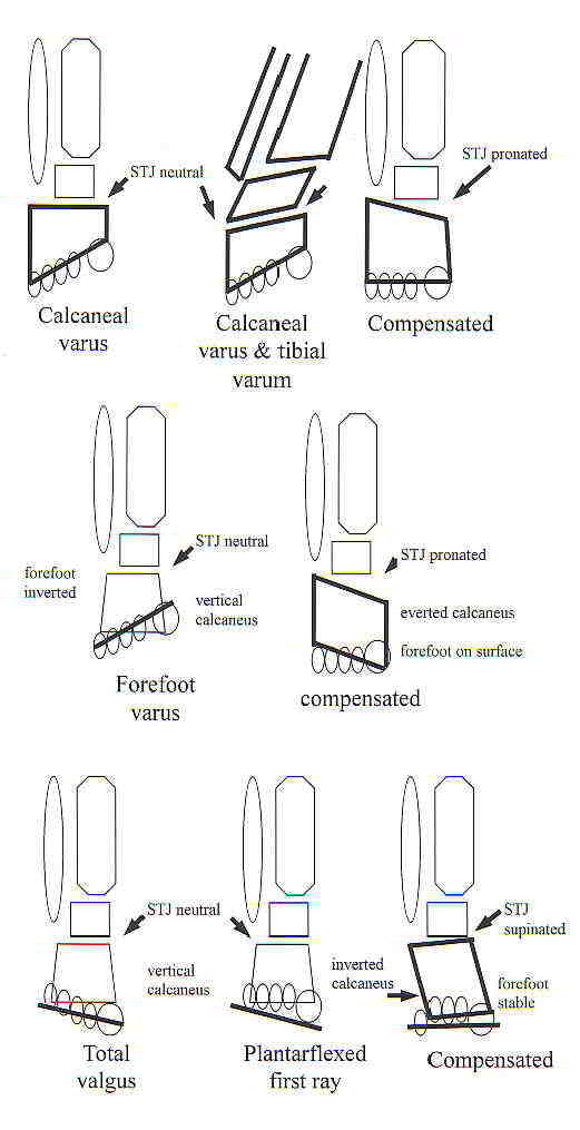
Orthotics have been advocated in recent decades for the correction of lower limb biomechanical anomalies (Landorf & Keenan 1998). Certain assumptions have been entertained, which stipulate that control over foot pronation reduces the susceptability to injury. Biomechanically, prolonged excessive pronation during the stance phase of gait requires additional internal rotation of the tibia resulting in increased kne flexion (McCulloch et al 1993). Notably, reductions in tibial internal rotation during gait have been demonstrated (Nawoczenski et al 1995). Importantly, changes to calcaneal inversion/eversion with the use of orthotics do not appear to be as significant as the reductions in tibial internal rotation with orthotic use (Nawoczenski et al 1995).
Alterations in the neuromechanical relationship throughout the entire limb (Hertel et al 2001) through changes in lower limb EMG activity (Nawosczenski & Ludewig 1999) and reflex potentiation (Blanpied et al 1995) could be important aspects of the stretch-shortening cycles of viscoelastic tissue which may be influenced by the altered lower limb biomechanics induced by orthotics. Additionally, improved synergistic action between muscles that could lead to load sharing, and therefore reductions in the percentage of maximal voluntary contraction (MVC) at which the muscles need to produce power was not established (Tomaro & Burdett 1993). Indeed the use of orthotics was shown to increase tibialis anterior activity (Tomaro & Burdett 1993). However, prolonged tibialis anterior activity above 85% MVC has been associated with the high number of injuries amongst runners as a result of early fatigue (Reber et al 1993). Interestingly, these latter authors also demonstrated increased peroneus brevis activity during increasing speeds with running, suggesting the need for increased lateral ankle stability. Interestingly, 1 hour of submaximal treadmill running with orthotics was shown to alter neuromuscular control by protecting against fatigue induced reductions in rapid force development characteristics of the plantar flexors (Kelly et al 2011, Med Sc Sp Ex, 43, 12, 2335-2343)
Orthotics are considered to support the medial longitudinal arch of the foot, thus relieving tension in the plantar fascia (Donatelli 1987) during stance phase of running. The plantar fascia is thought to be an extension of the triceps surae, which acts with a ‘windlass action' around the ankle to produce power and stability to the foot during running (Adelaar 1986). Thus, alterations in EMG activity in the medial and lateral gastrocnemius muscles may be expected, reflecting the need to improve both power output and joint stability with increasing speeds of running. Indeed, ankle and knee stability has been shown to increase through coactivation of opposing muscle groups as the speed of running increases (Kyroelaeinen et al 1999). Additionally, the hamstring muscles insert on the medial and lateral aspects of the lower leg and therefore are influenced by, or alternately influence, tibial rotation. Importantly, improved joint stability at the knee and ankle, through shoe & orthotic use, could result in reduced EMG activity for the same power output when compared to barefoot or shoe running. Moreover, the majority of power and energy were found to occur in the transverse and frontal planes, primarily associated with energy absorption controlling actions of the lower limb during the midstance and push off periods of gait (Sadeghi et al 2000). Therefore, if orthotics aid in controlling transverse and frontal plane motions then improved effciency of gait and running should occur.
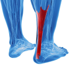
Interestingly, the posterior muscles of the lower limb show a uniform peak in activity during the midstance phase, as opposed to the terminal phase as was previously thought (Reber et al 1993). Their function appears to be to control ankle dorsiflexion through an eccentric contraction as the centre of gravity passes over the ankle (Reber et al 1993). Therefore, the biomechanical property of viscoelasticity in the series elastic components (SEC), contributing to the moments controlling ankle dorsiflexion (ie the plantar fascia and achilles tendons), may not be as significant since the SEC is shortening due to the lengthening of the contractile component (CC) of the biarticular gastrocnemius muscle. Yet during running the biarticular muscles (eg gastrocnemius) have been shown to perform concentrically, whereas the monoarticular muscles (eg soleus) first act eccentrically and then concentrically during the stance phase of running (Kyroelaeinen et al 1999). Clinically, anecdotal evidence suggests that orthotics with a heel raise/wedge could reduce the elongation not only of the achilles tendon, during stance, but probably of the plantar fascia as well. Notably, elongation and then shortening reactions are thought to impart force enhancement at the musculotendinous junction through the storage and release of elastic energy (Blanpied et al 1995). An analogy of the runner and the achilles tendon in particular resembling a pogo stick has been made, which describes this stretch – recoil cycle (Novacheck 1998). Orthotics have been shown to increase the amount of dorsiflexion and knee flexion during running (McCulloch et al 1993). Importantly, the duration of these actions increases during walking with orthotics, suggesting greater time for viscoelastic compliance.
Clearly, during running the viscoelastic compliance is likely to be reduced due to the increased velocities involved in the stretch-shortening cycle. Yet, the biarticular hamstring and gastrocnemius muscles are thought to have important eccentric – concentric functions, which aid efficiency through the storage and later return of elastic potential energy by the stretch of viscoelastic structures (Novacheck 1998). Additionally, efficiency is maintained through the transfer of energy from one body segment to another by the two joint muscles (Novacheck 1998). Importantly, unlike, walking where the interchange between potential and kinetic energy are out of phase, during running these are in phase (Novacheck 1998). Therefore, with increasing velocity of running, the hamstring muscles exhibit minimal change in length and hence mainly functions as an ‘energy strap' transfering energy from the tibia to the pelvis (Novacheck 1998).
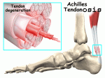
Anecdotally, orthotic modifications of footwear have been used clinically to treat achilles tendonopathy (Seggesser et al 1992). Theoretically, the effect of orthotics on achilles tendonopathy may be the result of reduced eversion (McCulloch et al 1993), improved talonavicular stability and thus improved ankle and knee stability (McCulloch et al 1993), resulting from reductions in tibial internal rotation (Cornwall & McPoil 1995, Eng & Pierrynowski 1994). Presumably, reduced midfoot pronation leads to reduced frontal plane ankle eversion could create less shearing across the achilles tendon. Additionally, reduced midfoot pronation may reduce transverse plane tibial internal rotation, thereby diminishing energy expenditure in non-sagital plane motions.
Saggital plane trunk flexion has a significant influence on hip and knee energetics during running. People who run in a more extended, backward leaning, posture exhibit higher energy generation and absorption at the knee extensors and lower energy generation in the hip extensors (Teng & Powers 2014, Med Sc Sp Ex, 47, 3, 625-630). Such postures would presumably also alter the weight bearing characteristics of the foot.
Clinically, soft 'off-the-shelf' orthotics have been shown to improve anterior knee pain (patellofemoral pain) in people with reduced ROM of dorsiflexion, low levels of pain (<2.2 on VAS), and who report immediate reduction in pain during lunging (Barton et al 2011). Improved synergistic muscle sharing between the medial and lateral gastrocs and the hamstring muscles can be expected, if enhance frontal plane motion can occur through a reduction in transverse and frontal plane motions. Alternatively, the frontal and transverse plane motions may be a necessary ‘damping' moment to reduce the likelihood of injury due to excessive rapid transfer of energy into viscoelastic structures controlling saggital plane propulsion during running. Therefore, it is hypothesised that orthotic use will decrease frontal and transverse plane motions in addition to enhancing EMG activity for frontal plane motion.
Methodology
A male subject was prepared for a 4 test protocol where running was compared over 4 conditions : barefoot, shoe running, shoe + orthotic running, shoe + orthotic + fast running.
Eight video cameras using strobed lights at 120 per secs, a Kistler force plate, and EMG data were used to make comparisons between the 4 conditions. Video equipment was calibrated for 90 seconds using a 499.93mm wand with 0.1 mm errors. The calcaneal angle was calibrated at 7 degrees. The force platform was calibrated for the subjects weight. The EMG was tested for impedence using an ampmeter where 10k W was excepted as normal. Gain settings using a power pack for telemetry sampling at a 400Hz frequency was considered satisfactory for ‘on/off' measures.
The skin was shaved, washed, and abraded for the application of surface EMG electrodes. Surface electrodes were placed at the medial and lateral gastrocnemius and hamstring muscles. Additionally, the subject had markers placed on the big toe nail, 1 st metatarsal head, 5 th metatarsal head navicular, lateral and medial malleolus, medial and lateral calcaneus, posterior calcaneus wand, medial and lateral condyles of the knee, mid tibia, mid thigh, and ASIS. The calcaneal wand was placed through a special window constructed through the sports shoes. The foot markers were removed from the foot surface for the shoe conditions.
The Kintrak project read the data, the segments were identified, the variables were created, the tables were created and the trials were applied. The data was inputed into the Matlab where the EMG was rectified and smoothed for linear envelope representation. The sampling frequency of data was changed to 360 per second. Each condition was repeated three times after a practice trial was performed.
Results & Discussion
The results of the 4 conditions for variations of angles are shown below.
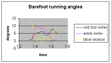
Fig 1.1 Barefoot running angles : external rotation of the tibia at heel strike is followed by an oscillation from internal to external rotation during the stance phase. Both the midfoot and ankle begin in inversion, but evert rapidly, before inverting again at push off.
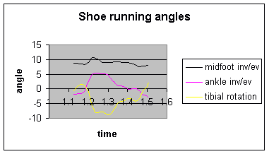
Fig 1.2 Shoe running angles : little external rotation of the tibia is followed by internal rotation during midstance and then progressive external rotation. The midfoot demonstrates little variability whereas the ankle inversion, followed by eversion and subsequent inversion at push offt.
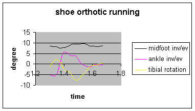
Fig 1.3 Shoe & orthotic running : little external rotation of the tibia is followed by minor oscillations into internal rotation, with subsequent neutral rotation at push off. The midfoot shows little variability, whereas the ankle deomnstrates inversion at heel strike, eversion in midstance and neutral position at pushoff.
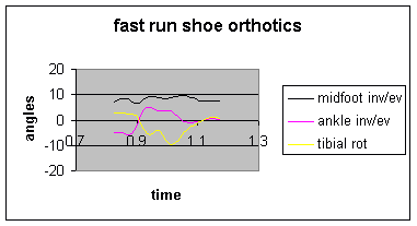
Fig 1.4 Shoe & orthotic fast running : slight external rotation of the tibia is followed by large oscillations into internal rotation with subsequent neutral position at push off. The midfoot shows little variability, whereas tha ankle is in inversion at heel strike followed by eversion at midstance and neutral position at push off.
The data suggests that the barefoot condition demonstrates the greatest variability of knee rotation and ankle inversion/eversion angles during running. The orthotics conditions quite clearly demonstrated neutral ankle and knee positions during the power generation phases of push-off. Interestingly, the shoe condition demonstrated a markedly longer period of stance, whereas the barefoot condition was slightly longer than the two orthotics conditions. Surprisingly little difference in the duration of stance was demonstrated between the fast run and slow run conditions. However, this may be explained by the power generating capacity of the stance phase and perhaps shorter flight phases in the fast run condition.
The surface EMG results for the 4 conditions follow.
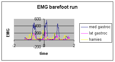
Fig 2.1 Surface EMG of medial and lateral gastrocnemius and hamstring muscles during barefoot running : Clear spikes of medial gastrocnemius muscle activity can be seen to be much greater than the spikes in lateral gastrocnemius and hamstring muscles.
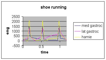
Fig 2.2 Surface EMG of medial and lateral gastrocnemius and hamstring muscles during shoe running : Greater synchronization of EMG activity was demonstrated, however the hamstring muscles showed the greatest amount of activity.
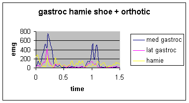
Figure 2.3 Surface EMG of medial and lateral gastrocnemius and hamstring muscles during shoe and orthotic running : medial gastrocnemius muscle showed the greatest amount of activity. Interesting, the hamstring muscles showed very little activity.
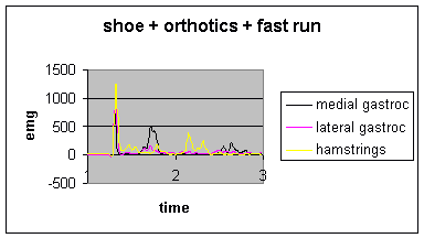
Fig 2.4 Surface EMG of medial and lateral gastrocnemius and hamstring muscles during fast running with orthotics : note the synchronization of spikes of activity between the three muscles at heel strike, followed by moderate asynchronous activity of medial gastrocnemius and hamstring muscles.
The EMG data is remarkable for the spikes of activity in the barefoot running condition. Particularly the medial gastrocnemius muscle had double the activity of the lateral gastrocnemius and hamstring muscles. When compared to the angular data, this may be interpreted as muscle activity controlling the angular pertubations of movement at the midfoot, ankle and knee. Strikingly, the shoe running condition demonstrated extremely large EMG values for all three muscles, especially when comparing these results to those of the other conditions. Explanations for such a large amount of EMG activity may reside in the enhanced neuromuscular stability required for the soft nature of the shoes without orthotics. Clearly, a lot of EMG activity appears to be wasted suggesting the possibility of early onset of fatigue with cummulative loading. All EMG activity is markedly reduced in the running condition with the orthotic compared to the condition without the orthotics. However, medial gastrocnemius muscle activity remains high, whereas lateral gastrocnemius activity is reduced when compared with the barefoot condition. Fast running demonstrates a synchronous spike nof activity at heel strike which may reflect the decelerating function of these muscles. The secondary hamstring muscle spikes may reflect the momentary reversal of tibial rotation and ankle inversion/eversion seen in the kinematic data. Additionally, the lack of further muscle activity and the minor spike of medial gastrocnemius may correlate with the oscillatory change from eversion to inversion seen in the kinematic data.
Bare Foot Running literature
Patellofemoral and ITB pain has been associated with increased hip adduction, hip internal rotation and reduced lumbopelvic rhythm. Investigations in 23 female habitually shod runners, comparing bare foot running with shod running, demonstrated reduced adduction, internal rotation and improved pelvic support, at initial contact and 10% stance, whilst running barefoot (MacCarthy C et al., 2015, Medicine & Science in Sports & Exercise. 47(5):1009-1016). When examining kinetic, kinematic and EMG data in AFL footballers whilst running on a treadmill in football boots (no arch support) versus running shoes with an average 10mm arch support, they found that in the latter condition there was an increase in knee, hip and ankle flexion resulting in a reduction in the peak torque thereby presumably reducing strain in the hamstring and calf muscles, which also correlated with reduced EMG activity. This may be significant in AFL where hamstring strains account for 15% of all injuries (Bartold SJ 2004, Medicine & Science in Sports & Exercise, 36(5), S167). Although a treadmill condition was used, investigators have found similar kinematic and kinetic data when comparing over gound to treadmill running provided the treadmill was reasonably stiff and speed was hel constant (Riley PO et al 2008, Medicine & Science in Sports & Exercise. 40(6):1093-1100).
. Metabolically, no differences were found during treadmill running, using 12 healthy male subjects who had extensive barefoot running experience using a light versus heavier shoe (Franz JR et al., 2012, Medicine & Science in Sports & Exercise. 44(8):1519-1525).
Running style may also influence the nature of the contrasting attitudes and experiences of runners and pratitioners to recommending various footwear. Some runners are natural heel - toe runners, others are forefoot runners which presumably places more load on the calf muscles, whilst others are mid-foot runners. Additionally, pose running has been introduced which is a novel running style in where the acromium, greater trochanter, and lateral malleolus are aligned in stance. When 20 heal-toe runners were intsructed in midfoot and pose running it was found that the latter was associated with shorter stride lengths, smaller vertical oscillations of the sacrum and left heel markers, a neutral ankle joint at initial contact, and lower eccentric work and power absorption at the knee than occurred in either midfoot or heel-toe running (Arendse RE et al 2004, Med. Sci. Sports Exerc., Vol. 36, No. 2, pp. 272–277).
Due to the contrasting nature of running barefoot, minimalist shoe running, shoe running and using orthotics it is suggested that care and precise diagnostic procedures and clinical reasons are needed when recommending any one of these strategies. In a study of 36 expereinced male runners over a 10 week period of transition to minimilist running shoes such as the VIBRAM Five Fingers an increase in bone marrow oedema in the foot was seen suggesting a slow transition is prudent when chaning to barefoot running style (Ridge ST et al., 2013, Medicine & Science in Sports & Exercise. 45(7):1363-1368). Clearly, the practitioner needs to advise their client of the gradual change to new loading, and also the alteration in running style from a more rear foot high loading style to a more mid - forefoot loading which may be accompanied by a reduction in stride length.
Injuries
Tendons act as buffers, delay energy absorption by muscles and therefore act as attenuators and shock absorbers and hence have implications for neural control. If tendons are injured, or if a pain state exists the ability to engage the elastic components of movement is diminished. In fact, people with chronic ankle instability have been shown to have decreased energy dissipation at the knee (Terada et al 2013, Med Sc Ex Sc, 44, 11, 2120-2128). Additionally, functional ankle instability has been associated with a deficit of ecccentric invertor rather than evertor strength as a result of too little ability to control lateral displacement of the shank over the foot (Munn J, 2003, Medicine & Science in Sports & Exercise. 35(2):245-250). This then leads to the question of whether or not orthotics aid or hinder eccentric invertor activity as reduced pronation would result in less tendon displacement. This may make those people also more likely to have injuries to the knee, hip and spine unless a precise professional guidance is followed.
Conclusions
The data demonstrates marked improvements in joint neutral positions when running using orthotics. Correlating the kinematic data with the EMG data appears to support the hypothesis that the use of orthotics may lead to more efficient running patterns, which in turn may reduce the susceptability to fatigue. Reduced fatigue may lead to reduced injuries through reduced purtubations of movement in the frontal and transverse planes. The data did not demonstrate any significant alterations in midfoot inversion/eversion in the three shoe running conditions. However, this may repesent sampling error, whereby the foot markers weren't as clearly marked as they were in the bare foot condition. Finally, the tibial rotation oscillation in the barefoot condition may reflect a somewhat externally rotated position of the tibia at heel strike and a stretch shortening cycle of various structures to bring about internal rotation of the tibia during midstance prior to external rotation at pushoff.
see also Inverse Dynamics of the ankle
Achilles Tendonosis (Pdf)
Last update : 3 September 2015


