Clinical reasoning for anterior hip pain
by Martin Krause
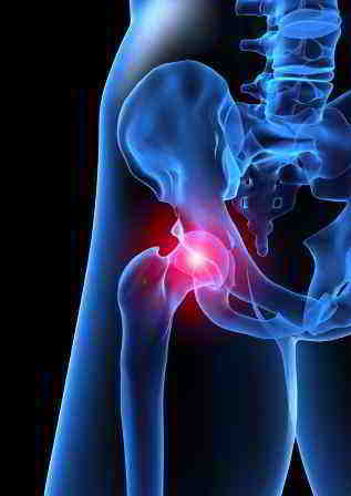
Anterior hip pain can result from various structures. The clinical reasoning process acts as a filter whereby many possibilities are reduced to the most probable. Frequently, inexperienced clinicians use little information to reach a diagnosis. In contrast, the experienced clinician uses multiple impairment variables to establish clinical patterns which should correlate with the events leading up to the pain as well as the consequences of the pain. By reducing the variables into pattern recognition the experienced clinician can use inductive & deductive reasoning to confirm their 'working hypothesis'. When the clinical features do not fit a known pattern, then deductive reasoning is used to examine the basics, correlate this with principles of patho-anatomy, biomechanics and neurophysiology to form a management strategy for the new clinical pattern. Importantly, the subjective examination and disability measures should correlate with the physical examinations impairment measures, which in turn should be used to assess the outcomes of treatment. In this manner the efficacy, and hence validity & reliability of each and every technique can be assessed. Traditional approaches to Maitland physiotherapy have discussed the importance of the reproduction of the pain. However, in the subacute and/or chronic scenarios any treatment which has an immediate effect on the impairment measure or the disability is considered a valid method of treatment.
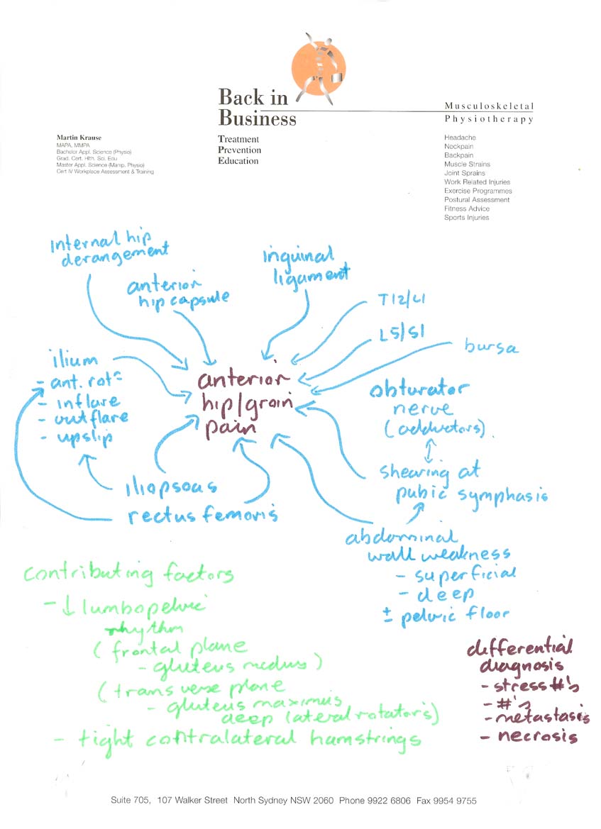
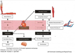
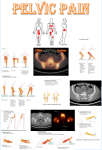
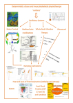
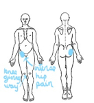
The following 'mind map' illustrates the multiple hypothesis which can be entertained when considering pain at the front of the hip.
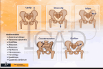
Hip - retroversion of the acetabulum
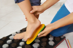
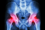
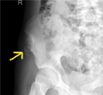
Common hip anommalies can be seen both clinically, as incresed range of motion of internal rotation, and the frequently seen bunions of the big toe. Further confirmation of hip retroversion can be seen using X-rays.
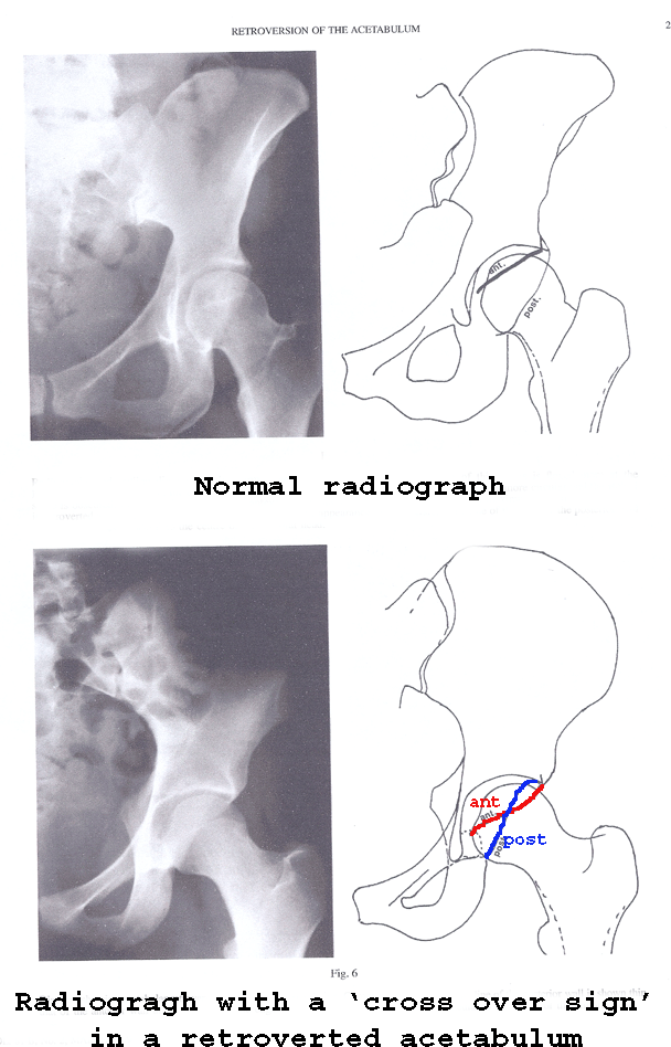
Reynolds, Lucas, Klaue (1999). Retroversion of the acetabulum - a cause of hip pain. JBJS, 81B, 2, 281-8
The retroversion of the acetabulum can lead to increased ROM of internal rotation with a concommittant loss of external rotation which in turn affects pelvic rotation during activities such as ambulation. Increased adductor muscle tone results in anterior translation of the hip and anterior rotation of the ilium (hemi-pelvis) which results in counter-nutation, and 'unlocking' of the SIJ. Torsion of the sacrum to the opposite side can place torsional load on the intervertebral disc between the fifth lumbar vertebrae and the sacrum.
Cam-pincer lesions
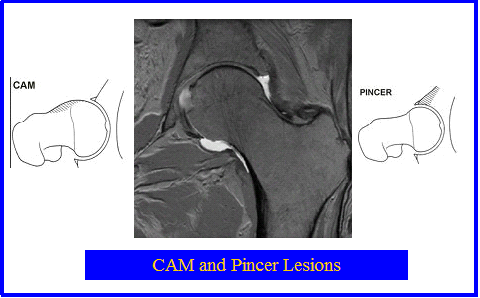
Cam and Pincer Lesions can occur in association with labral lesions. Excessive tension in the external hip rotators can cause anterior acetabular impingement. Iliopsoas tendonosis may be associated with anterior labral lesions. Excessive anterior rotation of the innominate can cause anterior hip impingement.
Pfirrmann et al (Radiology, 240, 3, 2006 pp778-785) used MRI measurements of alpha angles and the depth of the acetabulum to determine the risk and incidence of CAM and Pincer lesions in the hip. They concluded that a deep acetabulum and posteroinferior acetabular cartilage lesions were a characteristic finding of pincer impingement.
Lateral hip pain
Lateral hip pain can also occur with a retroverted hip. The external rotators such as obturator internus has a continuous myofascial membrane with the pelvic floor muscles. Strain on the external rotators such as the obturator internus can result in obturator nerve irritation causing increased adductor muscle tone, which requires a balancing contraction from the gluteus medius. Optimal gluteus medius contraction usually requires optimal internal pelvic muscles such as the horizontal fibres of the internal oblique and transverse abdominis.
Clinical reasoning
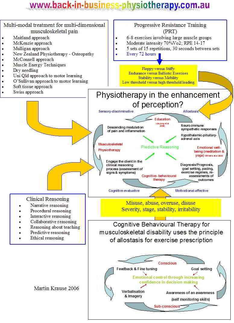
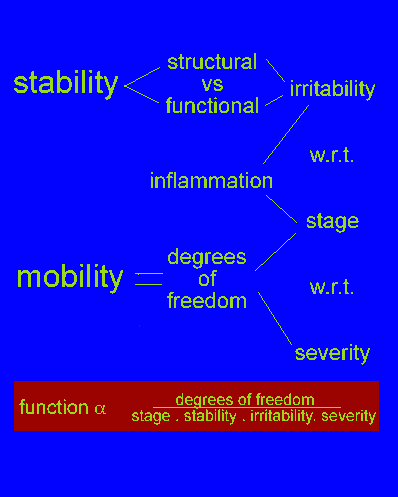
Clinical reasoning involves a process championed by physiotherapists whereby 'multiple hypotheses' are entertained and these are filtered down to the most plausible and probable.
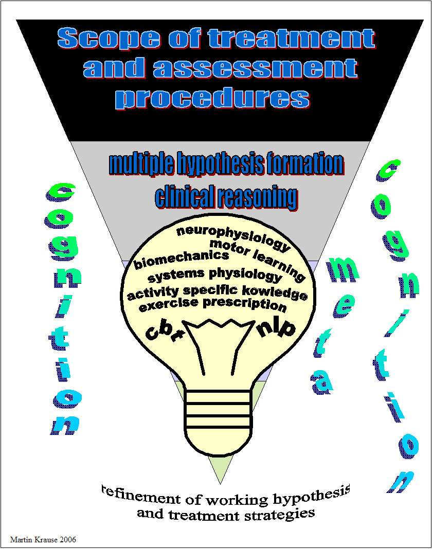
The filtering process uses a structured approach to questioning and hence examination, which more clearly defines the structure, or more frequently structures, involved in the on-going pathology, impairment and/or disability.
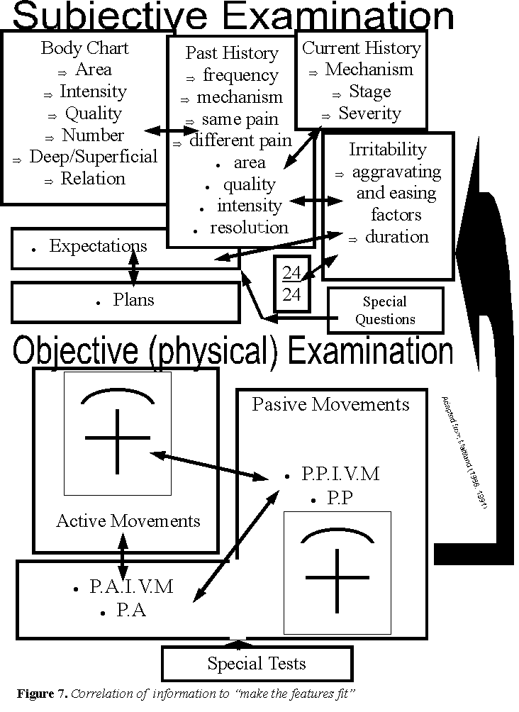
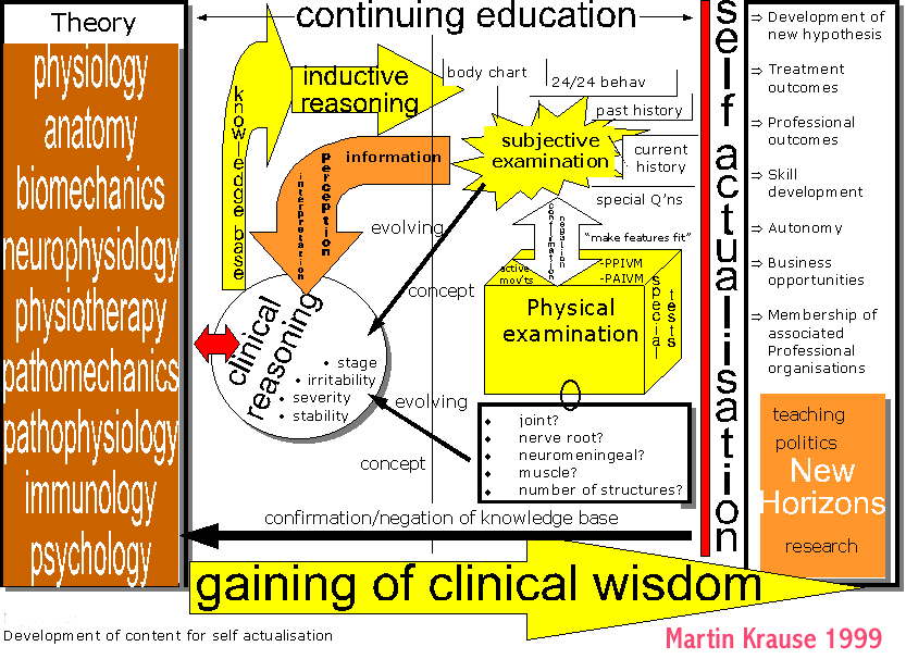
The structured reasoning approach allows the physiotherapist to evolve their clinical skills, particularly at a cognitive level to improve the selection of treatment techniques as well as provide a more confident prediction on the timeframe of management based on diagnosis and prognosis. Professional development through enhanced clinical outcomes strengthens the profession of physiotherapy, thereby averting an 'existential crisis'.
Ideally, the condition should be defined by the following categories. This will further help define the goals of treatment.
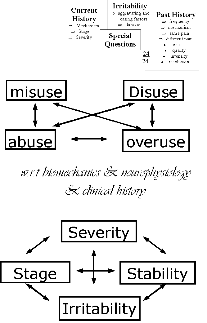
By defining these categories, the physiotherapist can gain confidence that they are not just relying on routines, but are basing their clinical judgement on reflective thinking.
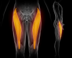
Increased hip adductor muscle tone
This video describes the basic tenate of increased adductor muscle tone leading to torsion in the intervertevral disc due to counter-nutation 'unlocking' of the sacroiliac joint (SIJ).
Increased muscle tension in the adductor muscles can result in an anterior shift of the head of the femur, which is countered by increased iliacus activity resulting in anterior rotation of the innominate, with consequent counternutation and instability of the SIJ.
Such increase in adductor muscle tension may also be accompanied by external oblique overactivity which can be assessed using the Active SLR, with varying emphasis on abdominal muscles, anterior and posterior compression of the pelvis and thorax ring relocations (see Muscle Energy Techniques {MET} section of this website). In the case of increased external oblique activity, the myofascia may be reactive and could respond to trigger point massage and myofascial releases as well as motor control strategies emphasising synergy with other muscles as well as the relaxtion phase of external oblique activity (Relaxation With Awareness {RWA}).
Abductor muscles
Gluteal tendonopathy is a common finding amongst young and old. It commonly occurs in conjunction with weakness across the core stabilisers of the abdominal and pelvic region. The gluteal muscles are important stabilisers against counternutation of the ilium (hemi pelvis) and work in conjunction with the contralateral abdominal, quadratus lumborum and latissiumus dorsi muscle in controlling lateral pelvic tilt. Interestingly, people presenting with gluteal tendonopathy demonstrate abductor strength deficits on both sides (Allison et al 2016, Med Sc Sp Ex, 48, 3, 346-352), suggesting weakness preceding and being the cause of clinical symptoms. However, we do know (from research on shoulders) that many tendonopathies demonstrate an immune-metabolic involvement, including infiltration of immune and fatty substances, both in the tendon as well as the adjacent bursa. Regardless, of cause or effect in any hip, pathology, the gluteal muscles must assessed for pelvic stabilisation deficiencies.
Treatment options include
- muscle energy techniques to the hip, ilium, hamstrings, rectus femoris and lumbar spine
- joint mobilisations to the lumbar spine and hip
- soft tissue and dry needling techniques
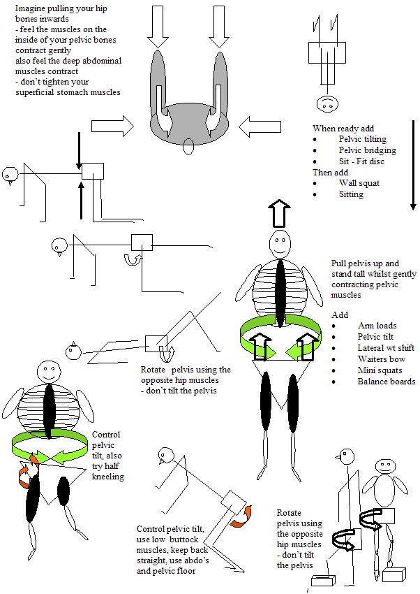
- exercises for deep hip rotator stability
- exercises for lumbo-pelvic rhythm
- stretches for the Psoas Major & Rectus femoris
- strengthening of the low, medium and high threshold lumbo-pelvic-hip stabilizers (dynamic & static)
- motor control strategies across hip-pelvis-lumbar-thorax
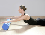
- integration of exercises into A.D.L. and perhaps a gym, yoga and/or physiocise programme
Engaging the client in the clinical reasoning process
Through the clinical reasoning process the physiotherapist engages the client in their healing process by helping their cognitive processes to refine and filter confusing and conflicting information (e.g. referred pain, mal-aligned ilia, etc)
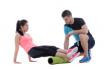
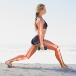
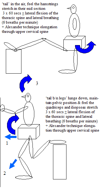
Stretches of the hamstrings and quadriceps whilst considering the position of the pelvis
The diaphragm can place an integral role in the management of hip displasia
The diaphragm has connections with the Psoas Major. The stomach muscles also attach to the lower 6 ribs. Through lateral diaphragmatic breathing the Psoas Major and stomach muscles elongate. Additionally, lateral diaphragmatic movement results in raising of the pelvic floor which has a stabilising effect on the deep hip muscles.
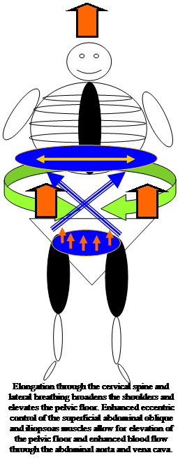
'Racking and stacking' of ribs may also be necessary to biomechanically optimise the tension placed on muscles and the ganglia of the sympathetic nervous system
Video demonstrating hip and thoracic stabilisation exercises
Conclusion
Treatment of the hip can be confusing due to the multiple structures which can play a role in hip pain. Importantly, this thorough examination process should be accompanied by a 'muscle energy' approach to the physical examination. Please view link to muscle energy techniques
Last update : 25 April 2016


